
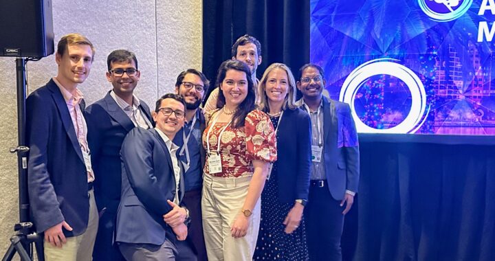
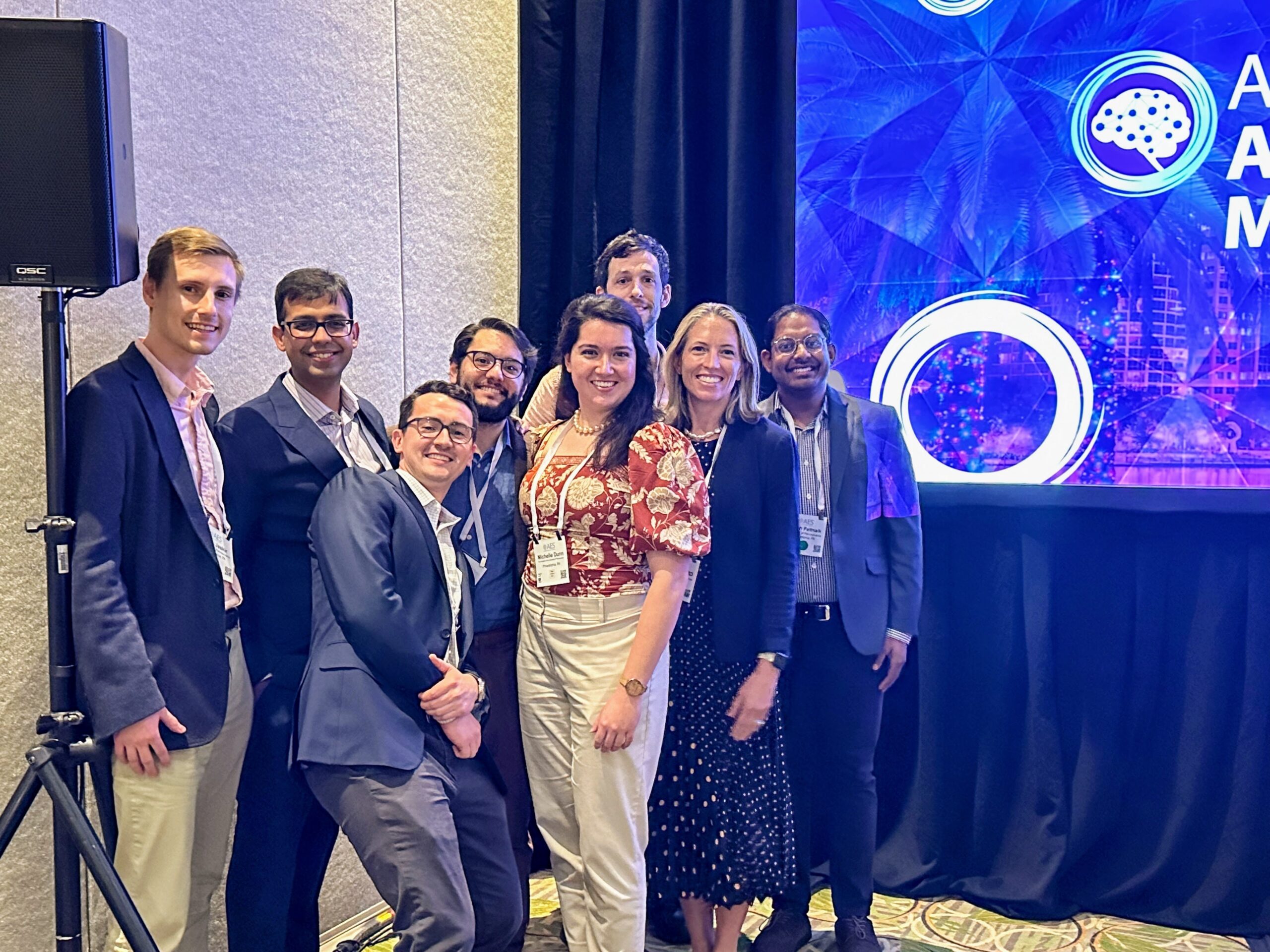
The Davis Lab presented many posters and talks at this year’s AES Annual Meeting in Orlando!
Talks
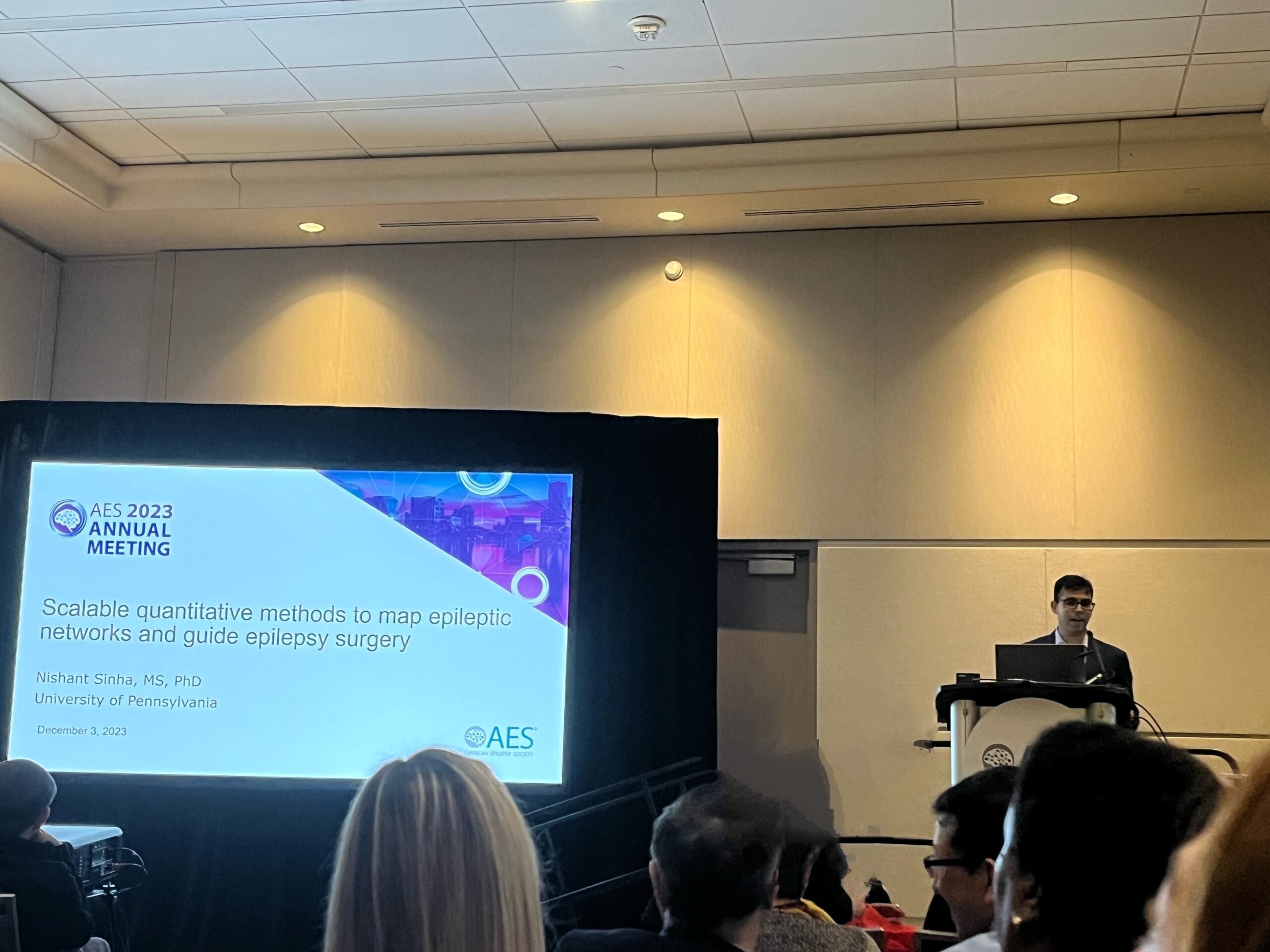
Nishant Sinha, PhD (Talk at SIG Neuroimaging Panel): Scalable quantitative methods to map epileptic networks and guide epilepsy surgery
Posters
Ryan Gallagher, MD student: Spatial Extent of Interictal Intracranial EEG Abnormalities Relates to the Focality of Epileptic Networks
Related paper: Quantifying interictal intracranial EEG to predict focal epilepsy.
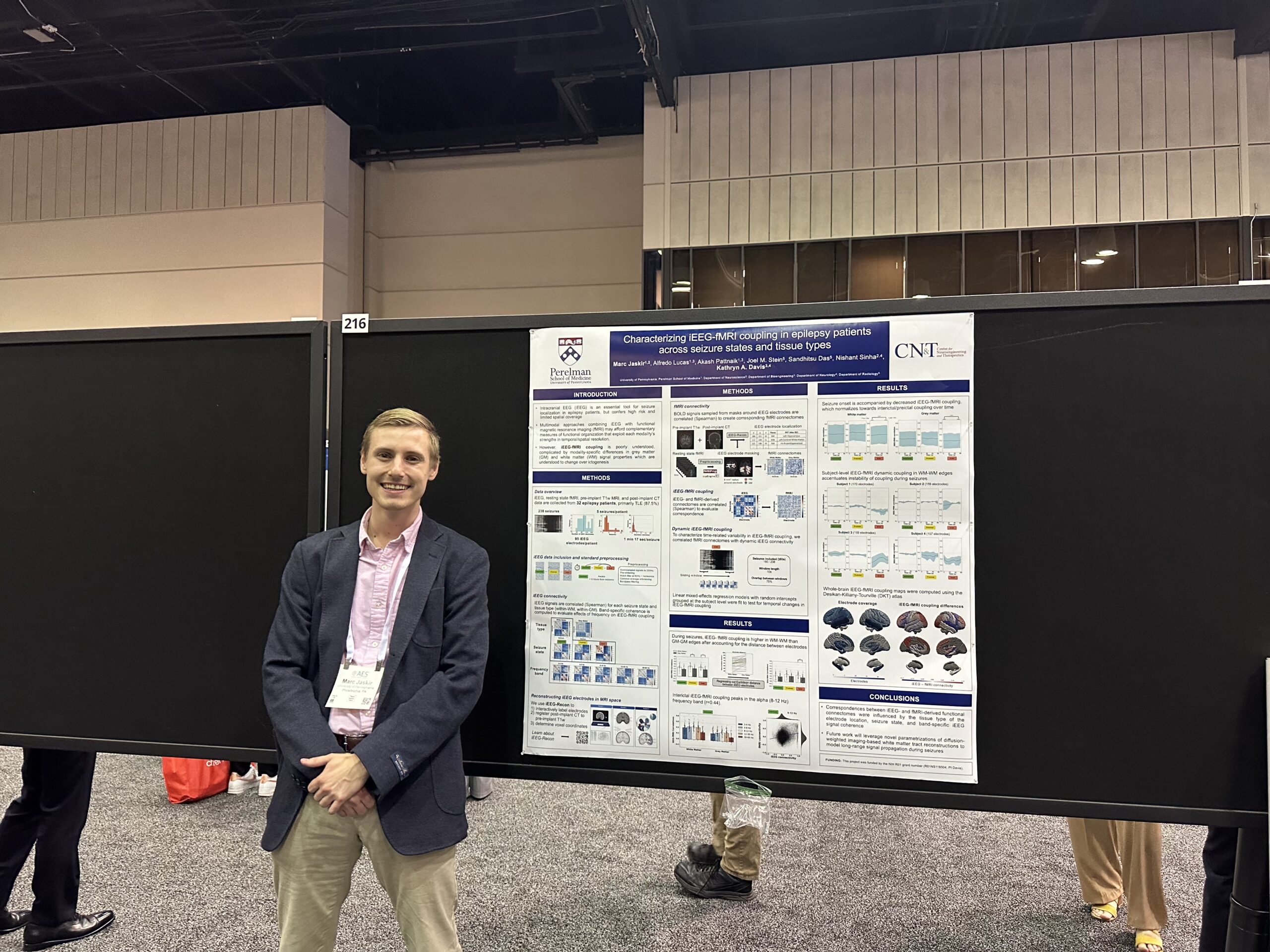 Marc Jaskir, PhD Student: Characterizing iEEG-fMRI Coupling in Epilepsy Patients Across Seizure States and Tissue Types
Rationale:
Intracranial EEG (iEEG) is an essential tool for seizure localization in epilepsy patients. However, iEEG is a highly invasive diagnostic procedure and its incomplete spatial coverage imposes limitations for whole-brain functional mapping approaches, which may reveal global network properties that are relevant to seizure onset and propagation. Multimodal approaches combining iEEG with functional magnetic resonance imaging (fMRI) may afford complementary measures of functional organization that exploit each modality’s strengths in temporal resolution and spatial coverage, respectively. However, iEEG-fMRI coupling is poorly understood1 and their relationship is further complicated by modality-specific differences between grey and white matter signal properties2,3 which are understood to change over ictogenesis. Therefore, we thoroughly characterized iEEG-fMRI coupling in epilepsy patients during different stages of ictogenesis and across different tissue types.
Methods:
Using resting state fMRI and interictal, preictal, and ictal iEEG data from 32 epilepsy patients, we evaluated differences in average iEEG-fMRI correlations and the associations between their graph theoretic properties (global efficiency) across different tissue types, seizure states, and frequency bands. We also used dynamic functional connectivity to elucidate seizure state-specific changes in iEEG-fMRI coupling over time.
Alfredo Lucas, MD/PhD Student:
Disparities and Algorithm Bias in Natural Language Processing of Epilepsy Outcomes
Exploring Cortico-cortical Evoked Potentials as a Marker of the Seizure Onset Zone
Guiding Intracranial EEG Implantation in Epilepsy Using Normative Brain Imaging
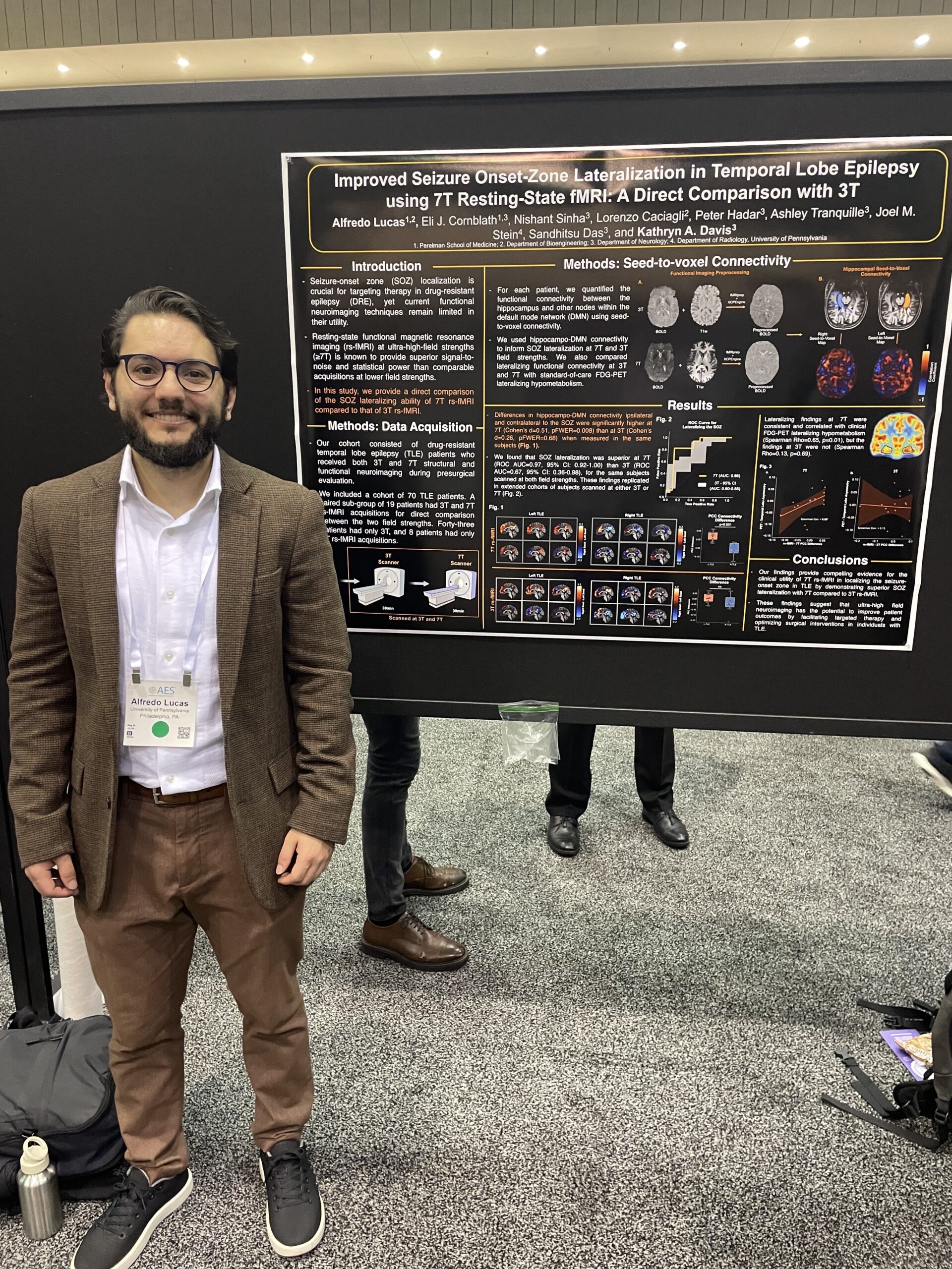
Improved Seizure Onset-Zone Lateralization in Temporal Lobe Epilepsy Using 7T Resting-State fMRI: A Direct Comparison with 3T
Multidien Cycles in Interictal Background Features of Long-Term EEG Recordings
Quantitative Intracranial EEG Abnormalities Across States of Consciousness in Drug-resistant Epilepsy
Role of Morphology in Interictal Spikes to Elucidate Seizure Generation
iEEG-recon: A Fast and Scalable Pipeline for Accurate Reconstruction of Intracranial Electrodes and Implantable Devices
Related papers:
Resting state functional connectivity demonstrates increased segregation in bilateral temporal lobe epilepsy
Dan Zhou, MD:
MEG Slow Wave Activity Associated with Brain Tumors in Patients with and Without Epilepsy
Rationale:
Epilepsy is prevalent in nearly half of patients with primary brain tumors. Recent evidence suggests that brain tumors can generate abnormal electrical activity in peritumoral tissue and affect local brain networks. Magnetoencephalography (MEG), a noninvasive imaging tool used for the functional mapping of eloquent cortex and localization of epileptogenic brain regions, has been used for cortical mapping prior to resective surgery. However, the evaluation of spontaneous interictal activity is often omitted. We aimed to characterize the interictal activity, including epileptiform discharges and slow activity, of patients who received presurgical MEG and compare the results between patients with and without brain tumor-related epilepsy (BTRE).
Methods:
We performed a retrospective analysis of the spontaneous interictal recordings in MEG during awake and sleep states in patients with primary gliomas who were recorded at the University of Nebraska Medical Center from July 2021 to March 2023. Focal slow wave activity (delta-theta band) was identified and processed for source localization using the equivalent current dipole method with Neuromag and Curry software. Dipoles were accepted when the goodness of fit was ≥80% and superimposed onto the individual patient’s brain MRI. A cluster was formed when ≥5 dipoles were localized closely together. Pre- and post-operative brain MRIs were used to determine tumor and resection sizes and locations, and the dipoles were then localized against the tumor resection sites.